0
My cartBasic Anatomy of the Eye
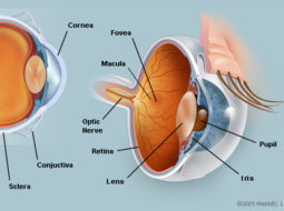
In this diagram, it can be seen that the eye can be considered as a ball or globe, with a transparent area (Cornea) and light entrance control (Iris and Pupil) at the front. The globe can be seen to comprise several layers.
The outermost layer is referred to as the Sclera, what you might call the “whites of the eyes”. The Sclera is actually covered in a thin mucous cell layer known as the Conjunctiva, which we shall discuss later. Beneath the Sclera is a blood vessel known as the Choroid, and beneath this is the Retina.
At the back of the eye, the Optic Nerve leaves to pass back under and through the brain and into the Visual Cortex, an area of the brain responisble for processing the images that fall on the retina. We can also see some other structures. The Optic Disc is an area on the retina where the optic nerve joins the eye.
Behind the Iris and Pupil, is the Lens of the eye, which can change shape to focus on objects at different distances. The lens is controlled and regulated by the Ciliary Body.
The eyes sit in the orbits, and are moved by six Extra Ocular Muscles. Covering the eyes is a Tear Film, and this is regulated and partly produced by the eyelids. The eyelids themselves contain many structures, including glands and the eyelashes.
It is interesting to consider in more detail each of the structures highlighted
1 = The Cornea 2 = The Iris 3 = The Pupil 4 = The Lens 5 = The Conjunctiva 6 = The Choroid 7 = The Optic NerveThe Cornea
The Cornea is the main lens of the eye, providing two thirds of its focusing power. It is the transparent entrance to the eye, comprising five layers. The layers comprise cells, collagen and other fibers. The cornea has no direct blood supply except at the edges, otherwise it would not be transparent.
The cornea has five layers, these being:
- The Epithelium, a layer of cells usually three deep. The cells are closely connected to one another.
- Bowmans membrane, this is an elastic membrane acting as a basement membrane for the epithelium and as a boundary to infection.
- The Stroma. This makes up the main part of the cornea. It is made up of cells called Fibroblasts, Collagen and other fibres such as Elastin. The fibres are tightly packed parallel in bundles, each bundle overlaps another at an oblique angle. The result is the transparency.
- Descemets membrane is the boundary between the Stroma and the last layer.
- The Endothelium. This is a single layer of tightly arranged and connected cells that facilitate the absorption of nutrients from the Aqueous inside.
Covering the cornea is the tear film, and this is important for maintaining the comfort of the eye, a clear optical surface and is essential for the passage of oxygen into the cornea.
Iris and Pupil
The Iris is a muscular pigmented layer resting at the back of the Anterior Chamber, behind the cornea. The hole in the centre of the Iris is called the Pupil. The size of the pupil is  controlled by two sets of muscles within the Iris. In dark conditions, the pupil is enlarged, called Mydriasis, to allow more light into the eye, so we can see more in the dark. In light conditions, the pupil is reduced, called Miosis, to prevent too much light getting into the eye.
The chamber formed between the iris and the cornea is called the Anterior Chamber. Aqueous circulates from the Posterior Chamber, through the pupil and into the anterior chamber. The aqueous is a clear liquid which provides oxygen and nutrients to the internal surfaces and structures of the eye, mainly the corneal endothelium.
At the point the cornea and iris meet, there is a drainage network called the trabescular meshwork in the angle between them. Through which the aqueous is removed. A constant flow then occurs, old aqueous leaving at the angle, the new aqueous produced by the Ciliary Body. Later, you will see how Glaucoma can arise from aqueous pressure building up.
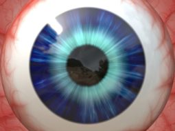
The varied amount of pigment and muscle fibres visible at the front of the iris give us the large variety of Iris colours we see in people’s eyes.
The Sclera
The sclera is commonly known as “the white of the eye”. It is the tough, opaque tissue that serves as the eye’s protective outer coat. Six tiny muscles connect to it around the eye and control the eye’s movements. The optic nerve is attached to the sclera at the very back of the eye.
The Conjunctiva
The conjunctiva is the thin, transparent tissue that covers the outer surface of the eye. It begins at the outer edge of the cornea, covering the visible part of the sclera, and lining the inside of the eyelids. It is nourished by tiny blood vessels that are nearly invisible to the naked eye. It provides oxygen to the cornea when we sleep and produces part of the tear film for the eye.
In older people, the conjunctiva can become thickened, raised and slightly yellow on either side of the cornea. These are termed Pingueclae ( singular Pingueculum ). They are harmless, and rarely need any treatment.
The conjunctiva can sometimes become inflamed and red, know as Conjunctivitus. This can be caused by bacteria, viruses, allergic reactions or by other eye problems.
The Choroid
The choroid is a rich vascular (blood vessel) layer lying beneath the sclera. It provides nutrients and exygen via the blood to the retina and the other internal eye structures. It also provides a support for the retinal cells to rest on and connect to.
The choroid is also rich in pigment, and this is essential for creating a boundary to light that could pass through the sclera, or bounce back from the back of the retina. The choroid, along with the iris and the ciliary body, forms an internal pigmented layer known as the Uvea.
The Retina
The retina is a multi-layered sensory tissue that lines the back of the eye, resting on the choroids. It contains millions of photoreceptors that capture light rays and convert them into electrical impulses. These impulses travel along the optic nerve to the brain where they are turned into images.
There are two types of photoreceptors in the retina: rods and cones.
The retina contains approximately 6 million cones. The cones are mainly contained in the Macula, the portion of the retina responsible for central vision. They are most densely packed within the fovea, the very centre portion of the macula. Cones function best in bright light and allow us to appreciate colour.
There are approximately 125 million rods. They are spread throughout the peripheral retina and function best in dim lighting. The rods are responsible for peripheral and night vision.
The picture shows a normal retina with blood vessels that branch from the optic nerve, cascading towards the macula.
The macula is the area we use most in our day to day vision. The detailed visual tasks we do, such as reading, depend on a healthy macula. There are many problems that affect the macula with age.
The Optic Nerve
The optic nerve is a bundle of nerve fibers (axons) that leaves the back of each eye and passes along what is known as the Visual Pathway to the back of the brain. This area of the brain is known as the Visual Cortex. Those areas associated with vision and processing what we see, make up almost an entire third of the brain!
Each optic nerve is packed with roughly a million axons, gathered from the nerve fiber layer of each retina. They gain what is called a myelin sheath as they pass out of the sclera at the back of each eye, doubling the diameter of the nerve. At this point, each optic nerve also gains a covering called the Meninges, which can be affected by Meningitis. So, it is important to remember that the optic nerve and the eyes are essentially part of the brain.
Blood vessels travel along the center of the optic nerve and into the eye, where they spread out all over the retina, apart from the macula, as can be seen in the diagram. These vessels are another source of oxygen and nutrients, and a channel for carbon dioxide and waste removal to many parts of the eye, including the retina. These vessels can be used to diagnose high blood pressure in the eye.
The optic nerve can lose nerve fibers, known as optic neuropathy. This can be caused by high pressure in the eye, leading to a condition known as Glaucoma.
The Lens
The lens lies behind the iris and pupil, and in front of the Vitreous. The ciliary body holds the lens in place via fibres known as Zonules. The lens is capable of changing the steepness of its front surface, and this allows the lens to change focus, a process known as accommodation.
The lens is layered rather like an onion, with numerous clear layers. The lens grows throughout life, and more and more layers are put down over the years. This means that the lens becomes thicker and stiffer, and loses its ability to change shape.
When this happens, it becomes increasingly more difficult to read, and we find it easier to push things further and further away to see them. Eventually, most people need reading glasses when this occurs.
This process of lens growth continues throughout life. Eventually, the older layers in the lens center get pushed together. This makes them very dense, and they become opaque. This forms what is known as a Cataract. Many of the residents in the homes we visit are elderly, and most of them will have some degree of cataracts, or will have had them removed.
It is clear from this fact, that even if someone has dementia, they will often benefit greatly from maintaining clear vision at all distances, including near.
The Ciliary Body
The ciliary body is part of the uvea. It consists of several parts, including the pigmented layer. Other parts of the ciliary body include the ciliary muscles, and the anchors to the lens zonules. At the back of the ciliary body, special cells produce and secrete the internal nutritional fluid called aqueous. The space made by the vitreous, the lens and the ciliary body is called the posterior chamber as mentioned earlier.
The ciliary body is responsible for many tasks, such as holding the lens in place, and helping control the lens shape for focusing, producing aqueous and as a route for blood vessels to other parts of the eye.
You can see from the diagram that behind the lens is another structure. This is the vitreous, and it is a clear gel like substance that fills up the inside of the eyeball, helping to hold the retina in place against the choroid. The gel is loosely attached at the optic disc and near the ciliary body all the way around the eye. Sometimes part of the vitreous can come away and cause symptoms of flashing lights and sudden severe floaters in the vision.


